
Schematic representation of the respiratory system. Download
The respiratory system aids the body in the exchange of gases between the air and blood, and between the blood and the body's billions of cells. It includes air passages, pulmonary vessels, the.
Respiratory system Canadian Lung Association
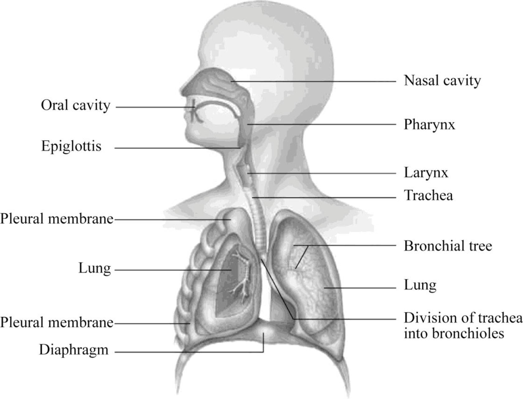
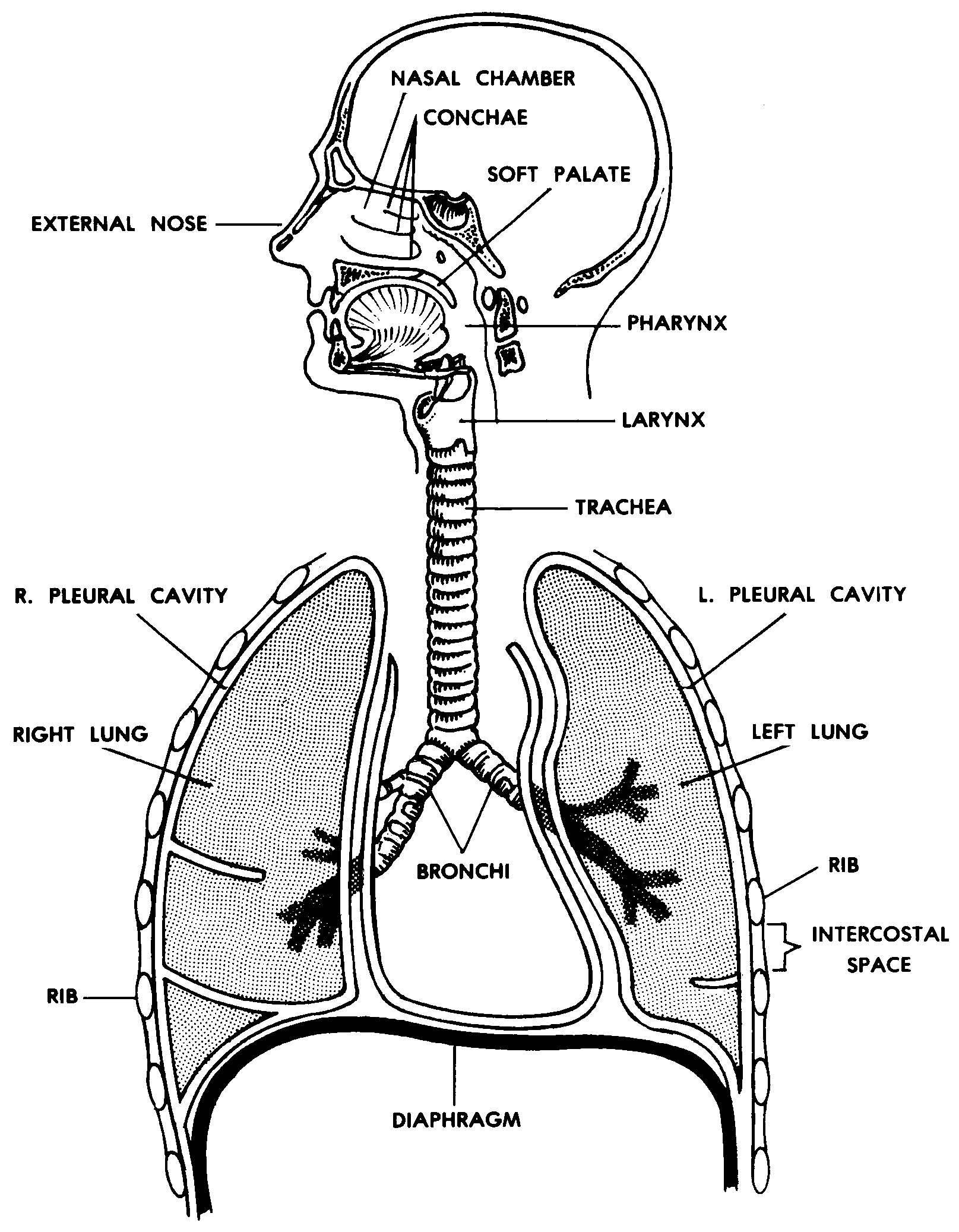
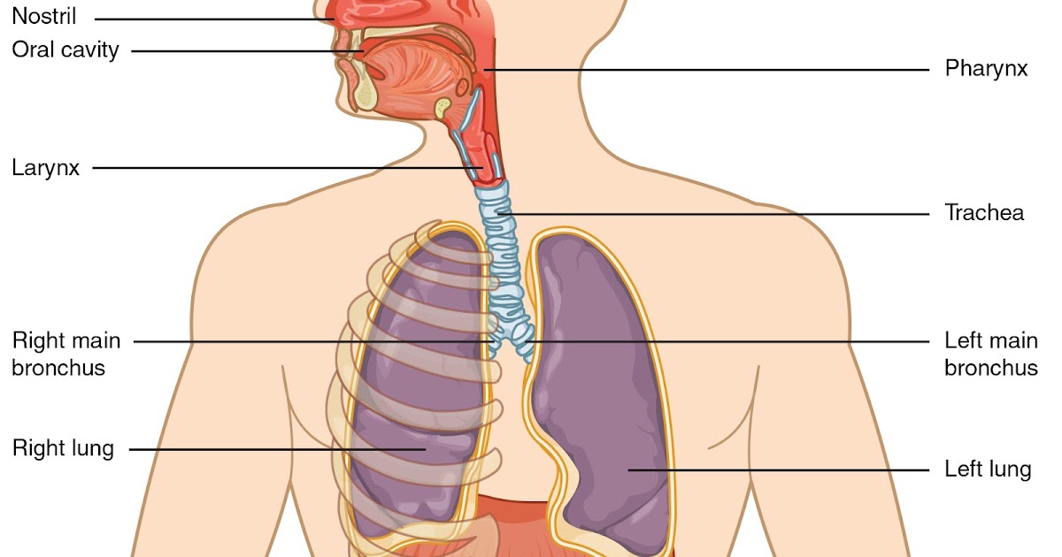
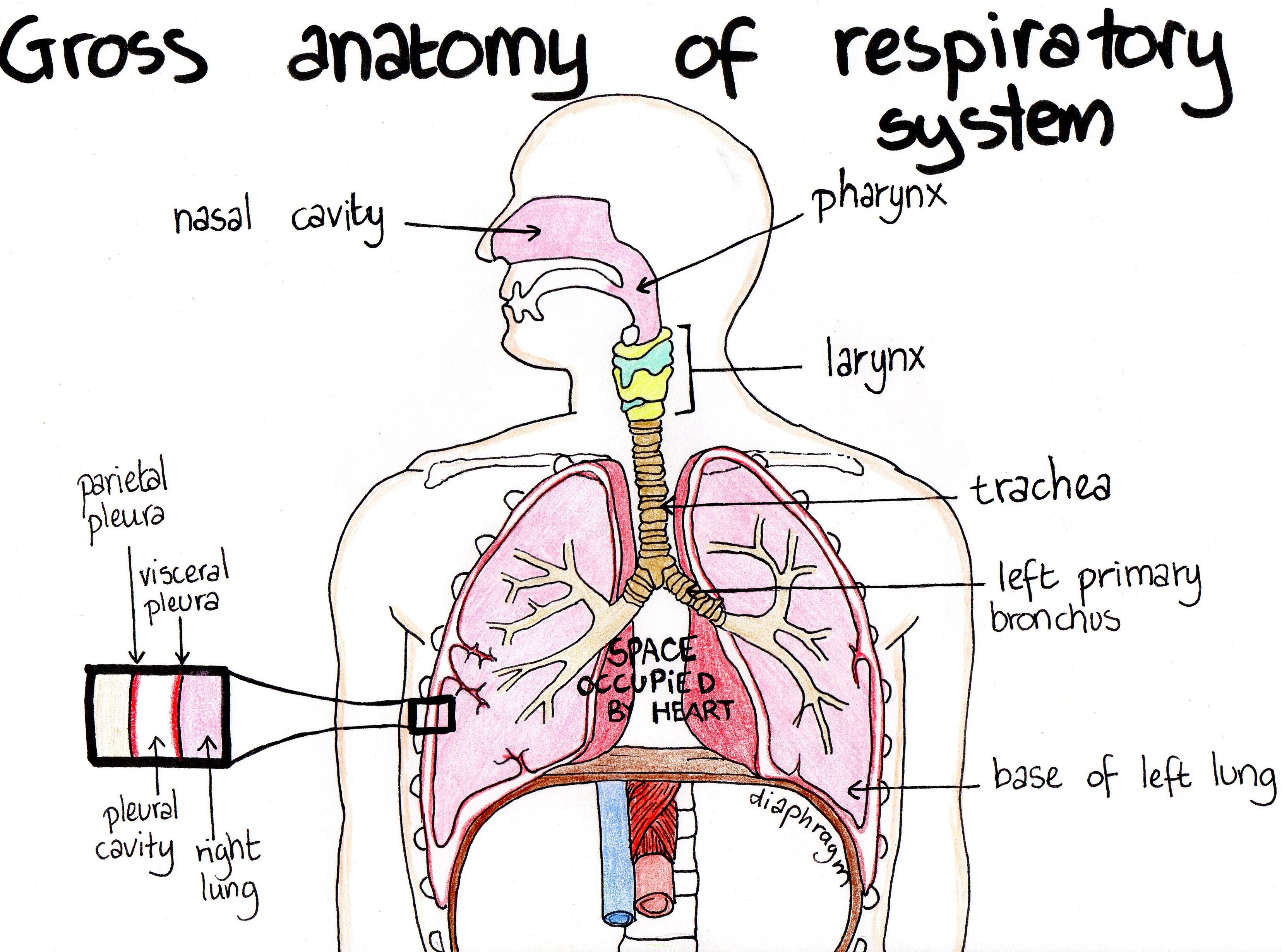
The organs of the respiratory system form a continuous system of passages called the respiratory tract, through which air flows into and out of the body. The respiratory tract has two major divisions: the upper respiratory tract and the lower respiratory tract. The organs in each division are shown in Figure 16.2.2 16.2.

THE RESPIRATORY SYSTEM Resources that may be used
Respiratory System Labeling Interactive — Quiz Information This is an online quiz called Respiratory System Labeling Interactive You can use it as Respiratory System Labeling Interactive practice, completely free to play.
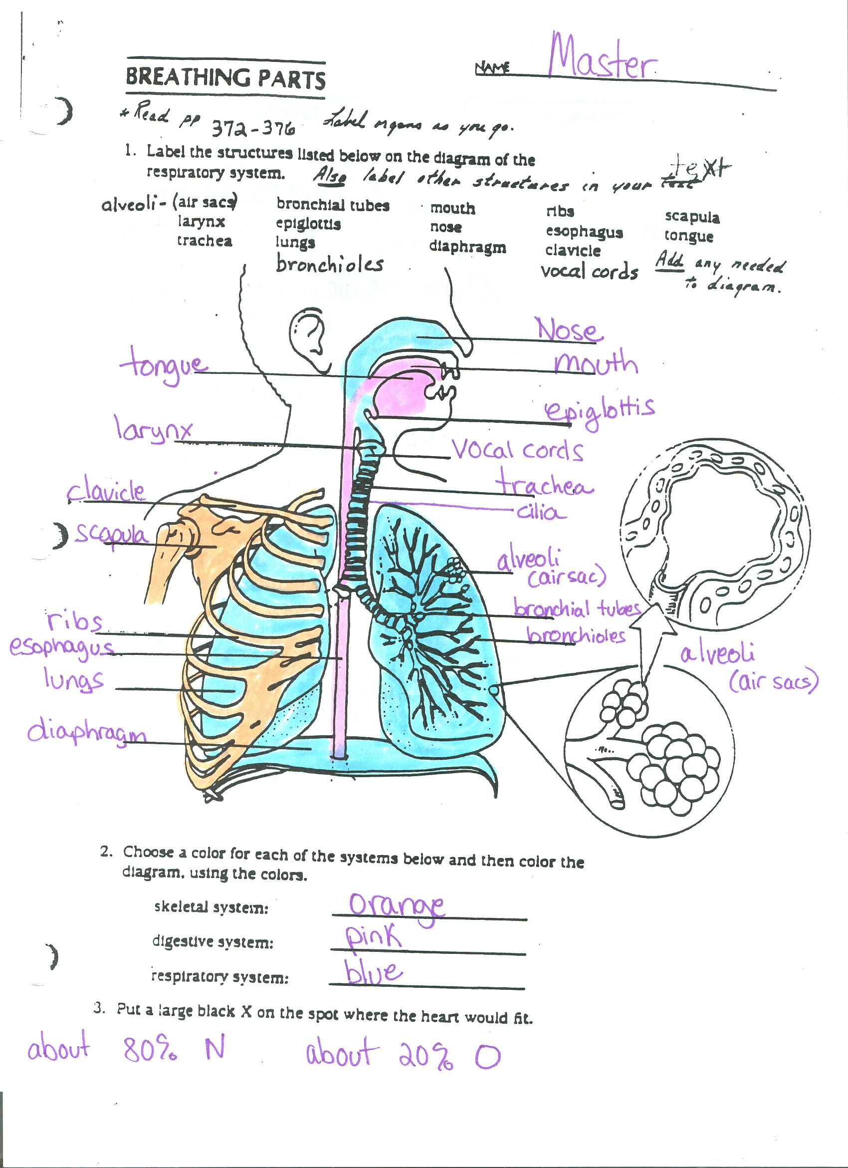
In the diagram below, label the parts of the respiratory system and the
Respiratory system interactive activities like this one are fantastic tools for helping your class learn about the human body! Interactive labelling activities are brilliant for introducing topics like this to a class as they encourage the visualisation of what's being taught, which can help kids understand these topics easier!
CLASS BLOG BIO 202 Respiratory System Worksheet
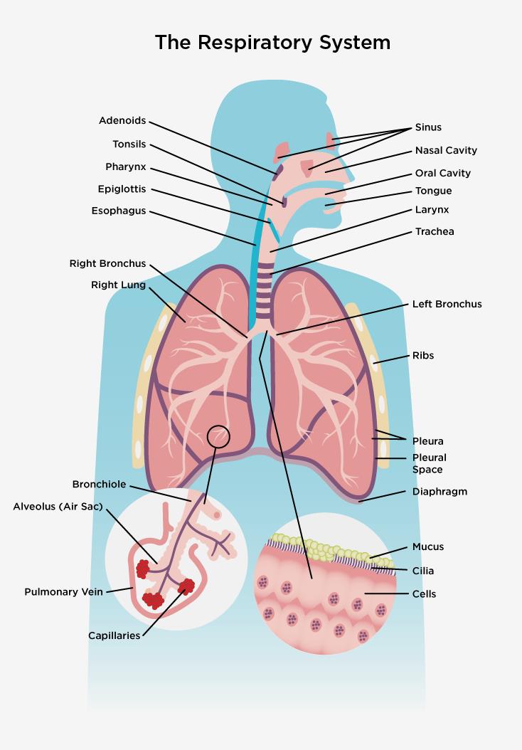
The cells of the human body require a constant stream of oxygen to stay alive. The respiratory system provides oxygen to the body's cells while removing carbon dioxide, a waste product that can be lethal if allowed to accumulate. There are 3 major parts of the respiratory system: the airway, the lungs, and the muscles of respiration.
Anatomy of the human respiratory system. (Reprinted with permission
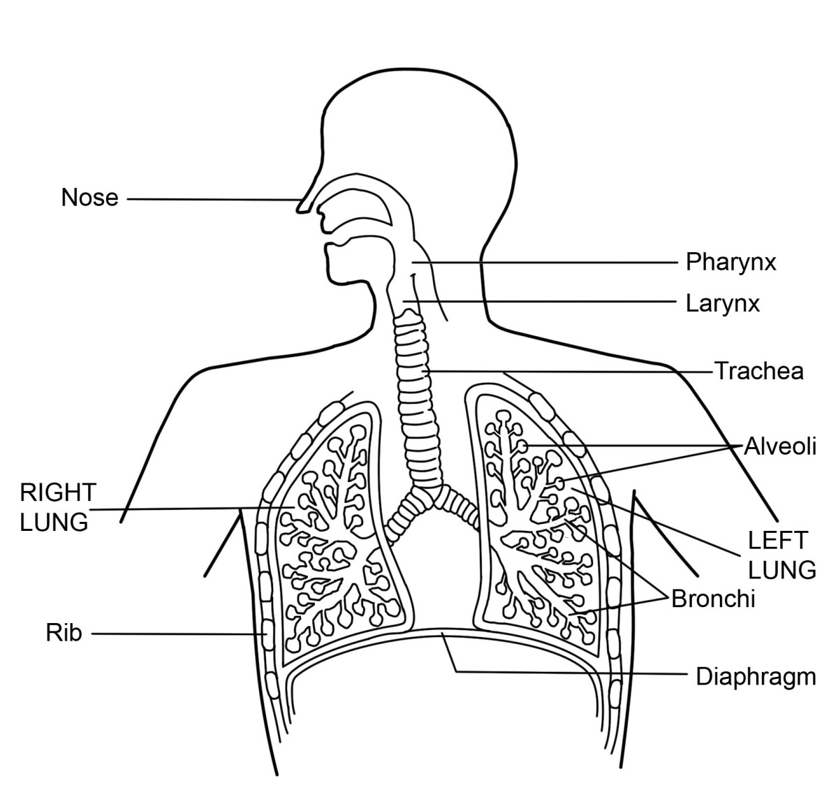
Alveoli are connected to their neighbors by alveolar pores, which help maintain equal air pressure throughout the alveoli and lung ( Figure 22.11 ). Figure 22.11 Structures of the Respiratory Zone (a) The alveolus is responsible for gas exchange. (b) A micrograph shows the alveolar structures within lung tissue.
Images 07. Respiratory System and Breathing Basic Human Anatomy
Strengthening Team-based Education in Practice Copyig Pappoaleaning 21 wwwepappoaleaningog PMD 1 Developed by Madeline Cox Page 1 of 2 Developed by Madeleine Cox

THE RESPIRATORY SYSTEM
The structures of the upper respiratory system, or respiratory tract, allow us to breathe and speak. The nose and nasal cavities provide airways for respiration. The paranasal sinuses surround the nasal cavities. The pharynx connects the nasal and oral cavities to the larynx and esophagus. The larynx and vocal cords allow us to breathe and talk.

Label the parts of your respiratory system Diagram Quizlet
Term. epiglottis. Definition. piece of cartilage that covers the larynx to prevent food from entering the airway. Location. Start studying respiratory system labeling. Learn vocabulary, terms, and more with flashcards, games, and other study tools.
Respiratory System Diagram and Vocabulary MS. CRAWLEY
Match each pair by dragging from rifgt to left. When complete choose the Check button.
RESPIRATORY SYSTEM
With a labeled diagram, you can see all of the main structures of an organ system together on one page - great for helping you to memorise the appearance of several structures and their relations. Unlabeled diagrams can then help you to put your memory to the test. Below, you'll find the respiratory system labeled and unlabeled on two.

Draw a diagram of respiratory system and label the following 1.parr
Respiratory system. The body system responsible for gas exchange between the body and the external environment. Pharynx (throat) Tube connected the nose/mouth to the esophagus. Larynx (voice box) Tube forming a passage between the pharynx and trachea. Trachea. Tube connecting the larynx to the bronchi of the lungs. Bronchi.

C.1. Introduction to the Respiratory System
View Original Image at Full Size. Labeled diagram of the lungs/respiratory system. Image 37789 is a 1125 by 1408 pixel WebP Uploaded: Jan10 14. Last Modified: 2014-01-10 12:15:34

Human Respiratory System 7 Download Scientific Diagram
The Nasal Cavity. The nasal epithelium (Figure 20.6.1 20.6. 1 is lined with ciliated pseudostratified epithelial with goblet cells and this makes up the mucosal layer. Deep to this layer will be numerous bipolar cell nuclei. You will also find Bowman's (olfactory) glands that secrete mucus to help lubricate the mucosal layer and to dissolve.
What is the Respiratory System Diagram and Function HubPages
Label the Respiratory System Structures ©Sheri Amsel www.exploringnature.org pleura pleura 1. 2. 3. 4. 5. 6. 7. 8. 9. 4 5 6 7
Respiratory System Medical Biology illustrated notes
eChalk HTML5 resource. www.eChalk.co.uk. Label the parts of the respiratory system. This resource also contains a diagram of alveoli. (worksheet)